Researchers Map Glycosylation Patterns Associated With Alzheimer’s Disease
Using new methodology, University of Kentucky researchers have mapped the variations in sugar chains attached to brain proteins from deceased healthy individuals or individuals with Alzheimer’s disease.
Thus far, no effective treatments for Alzheimer’s disease (AD) are available. New approaches to preventing the progression of this devastating neurological disease are desperately needed.
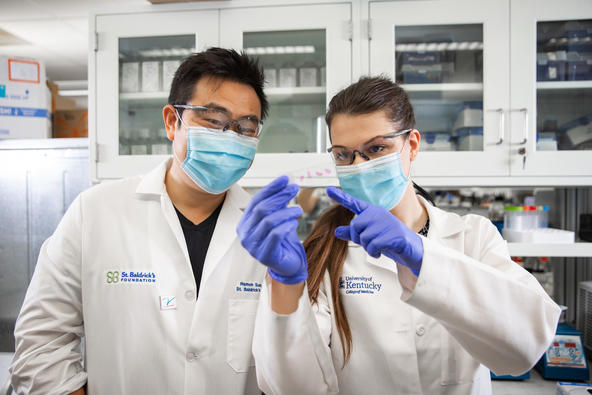
The laboratories of Ramon Sun, Ph.D., assistant professor of neuroscience in the UK College of Medicine and Markey Cancer Center researcher, and Matthew Gentry, Ph. D., professor of molecular and cellular biochemistry and director of the Lafora Epilepsy Cure Initiative, UK College of Medicine, developed a novel imaging method to identify the specific patterns of sugar molecules that are attached to proteins within a tissue. The form of sugar attachment that they studied is called N-glycosylation. They applied this methodology to analyze this “sugar code” in the brains of two mouse models of AD and in individuals who died from dementia.
The mouse models that they studied represent two different pathologies commonly found in AD patients. In one, the mice accumulate the protein Aβ (amyloid beta) in the brain; in the other, the mice accumulate abnormal forms of the protein tau in the brain. Despite having different underlying pathologies, both mouse models exhibited increased N-glycosylation in both the frontal cortex and the hippocampus.
They also analyzed samples from brains of three age-matched individuals and three patients with Aβ-type Alzheimer’s disease. Like the mouse brains, there was increased glycosylation in the frontal cortex region in the brains from AD patients. However, in contrast to what was observed in the mice, the hippocampus regions of the AD patients had reduced N-glycosylation.
The results define regionally specific differences between the frontal cortex and hippocampus in human AD patients and matched controls. Specifically, increased N-glycosylation was observed in regions of the frontal cortex in the AD brain and decreased N-glycosylation in the hippocampal regions. Furthermore, this study highlights a fundamental difference in N-linked protein glycosylation patterns in the hippocampal region between mouse models of AD and human patients.
“This study will potentially aid in the development of new research directions, new therapeutic targets, and biomarker evaluation for the future treatment and diagnosis of AD,” said Sun. This work was recently published in Alzheimer's & Dementia, the journal of the Alzheimer’s Association.
The importance of understanding N-glycosylation patterns and regulation of this biochemical process in the brain is the topic of a review in Trends in Endocrinology & Metabolism by Sun and Gentry and their teams. The biosynthesis of N-linked protein sugar chains is an under-studied branch of glucose metabolism. Within cells, glucose can be used for energy production or building complex chains of sugars that modify proteins or lipids. These processes compete for a finite source of glucose in cells.
“In the central nervous system, N-linked protein glycosylation is critical for both neurons and glial cells,” said Tara Hawkinson, a doctoral student in the College of Medicine and the lead author of this manuscript. This process controls many aspects of key proteins involved in neuronal activity. Aberrant glycosylation can drive dysfunction and death of neurons.
Consequently, altered protein glycosylation can contribute to various types of neuronal disorders, ranging from those associated with loss of neurons, such as Alzheimer’s disease and Parkinson’s disease, to those associated with defective neuronal connections, such as schizophrenia and developmental neurological disorders.
The methods developed by the Sun and Gentry laboratories for evaluating the spatial distribution of the sugar code of N-glycosylation patterns in the brain will enable researchers to ask key questions about how these patterns are altered in pathological conditions.
“With these methodological advances, we can begin to answer questions regarding how brain cells coordinate glucose metabolism to balance energy needs and glycosylation needs, how alterations in the sugar code contribute to neurological disorders and begin to develop therapies to address these issues,” said Gentry.
Research reported in this publication was supported by the National Institute on Aging of the National Institutes of Health under Award Number R01AG066653 and the National Institute of Neurological Disorders and Stroke of the National Institutes of Health under Award Number R35NS116824.
The content is solely the responsibility of the authors and does not necessarily represent the official views of the National Institutes of Health.

